

企业QQ

官方微信


Super Bright 600是串联染料,由Super Bright 436和受体染料串联而成。
►激发光:405nm
►发射光:600nm
►可替代染料:eVolve 605, Violet 605 (BV605
►检测滤片:610/20 bandpass
档案详细资料:
1. Super Bright 436 vs Brilliant Violet 605:亮度与BV421相当,尤其适合低丰度表达的抗原。
| Super Bright 600 antibody conjugates offer similar brightness to Brilliant Violet 605-conjugated antibodies.(A) Human peripheral blood cells were stained with Anti-CD3 (clone OKT3)conjugated to Super Bright 600 (red histogram) or Brilliant Violet 605 (grey histogram), using the same concentration of antibody. (B) Mouse splenocytes were stained with Anti-CD8a (clone 53-6.7) conjugated to Super Bright 600 (red histogram) or Brilliant Violet™ 605 (grey histogram), using the same concentration of antibody. | 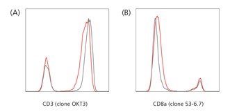 |
2. Super Bright 436 vs eVolve 605:亮度比eVolve 605要高。
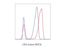 | Human peripheral blood lymphocytes were stained with Anti-CD3 (clone OKT3)conjugated to Super Bright 600 (red histogram) or eVolve 605 (blue histogram), using the same concentration of antibody. |
3. Super Bright 600光稳定性
(A) Mouse splenocytes were stained with Anti-CD4 (clone RM4-5) Super Bright 600and were either analyzed immediately (red histogram), left in the dark overnight (blue histogram), or left exposed to light overnight (green histogram). (B) Human peripheral blood cells were stained with Anti-CD8a (clone RPA-T8) Super Bright 600 and were either analyzed immediately (red histogram), left in the dark overnight (blue histogram), or left exposed to light overnight (green histogram). | 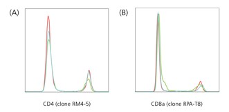 |
4. Super Bright 436 固定后的稳定性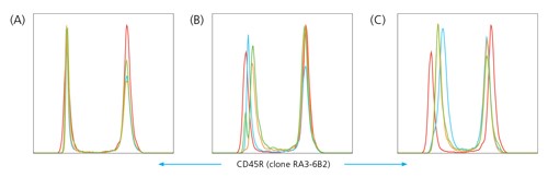
Stability studies indicate that Super Bright 600 exhibits a minimal loss of fluorescence when cells are exposed to various fixatives Mouse lymphocytes were stained with Anti-CD45R/B220 (clone RA3-6B2) Super Bright 600 and:
(A) were left unfixed (red histogram), or fixed in IC Fixation buffer for 30 minutes (blue histogram), 24 hours (orange histogram), or three days (green histogram), followed by a wash in Permeabilization buffer.
(B) were left unfixed (red histogram), or fixed in Foxp3/Transcription Factor/Permeabilization buffer for 30 minutes (blue histogram), 24 hours (orange histogram), or three days (green histogram), followed by a wash in Permeabilization buffer.
(C): were left unfixed (red histogram), or fixed in IC Fixation buffer followed by 90% methanol for 30 minutes (blue histogram), 24 hours (orange histogram), or three days (green histogram), followed by a Flow Cytometry Staining Buffer wash.
5. 多色分析示例- Super Bright 600.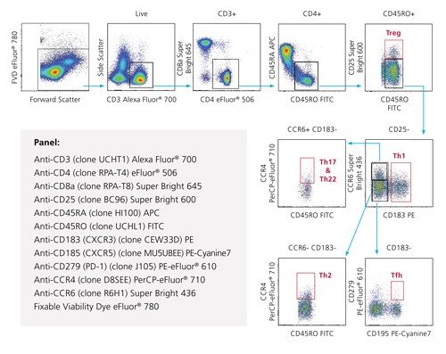
Normal human peripheral blood cells were aliquoted in the presence of Super Bright Staining Buffer (Cat. No. SB-4400), then surface stained with the indicated reagents. Viable cells, as determined by staining with Fixable Viability Dye eFluor® 780, were used for analysis to discriminate various T cell subsets.