企业QQ
官方微信

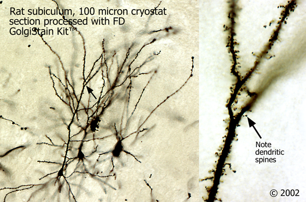
Golgi-Cox impregnation1, 2has been one of the most effective techniques for studying both the normal and abnormal morphology of neurons as well as glia. Using the Golgi technique, subtle morphological alterations in neuronal dendrites and dendritic spines have been discovered in the brains of animals treated with drugs as well as in the postmortem brains of patients with neurological diseases3, 4. However, the unreliability and the time-consuming process of Golgi staining have been major obstacles to the widespread application of this technique
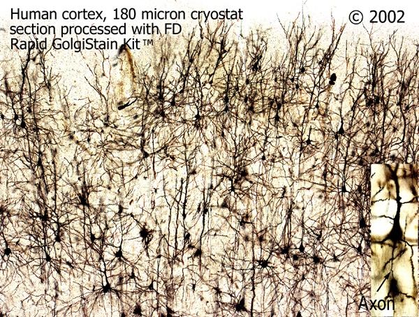
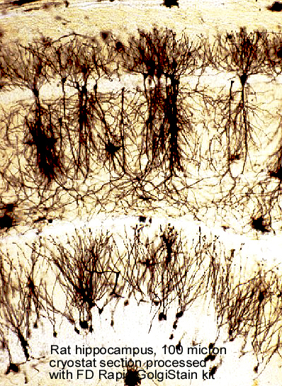
FD Rapid GolgiStain™ Kit is designed based on the principle of the methods described by Ramón- Moliner2, Glaser and Van der Loos5. This kit has not only dramatically improved and simplified the Golgi-Cox technique but has also proven to be extremely reliable and sensitive for demonstrating morphological details of neurons and glia, especially dendritic spines. The FD Rapid GolgiStain™ Kit has beentested extensively and widely usedon the brains from several species of animals as well as on the specimens of postmortem human brains.
  |  |
Store at room temperature
Solution A 125 mlSolution B 125 ml Solution C 125 ml x 2 Solution D 125 ml Solution E 125 ml Glass Specimen Retriever2Natural hair paintbrush2 Dropping bottle 1User Manual1
Materials required but not included:
References:
matriks/Shikari® (S-ATP) Anti-Pembrolizumab ELISA w/confirmation/PEM-QNS-KEY
intactgenomics/Methionine Auxotrophic LBA4404 | ig® | Intact Genomics/15x50μl/1076-15
goprolytix/Proteolytic Activation of Prothrombin/1 mg, 200 µg/BCT-DFP
novateinbio/Dynorphin A ( 13-17 ) Porcine Peptide/5mg/PE-54024_5mg
matriks/Shikari® (Q-ETA) Etanercept ELISA/ETA-FD-ENB
intactgenomics/Agrobacterium ElectroComponent Combo Pack | Intact Genomics/12x50µl/1290-24
Jackson/Peroxidase AffiniPure Goat Anti-Mouse IgG (H+L)/2.0 ml/115-035-003
Jackson/Peroxidase AffiniPure Goat Anti-Rabbit IgG (H+L)/2.0 ml/111-035-003
Qubit™ dsDNA BR Assay Kit Q32853现货
Invitrogen Q32855 Qubit™ RNA HS Assay Kit现货促销
LUCK-1G,D-Luciferin, Potassium Salt 钾盐现货
GoldBio/D-Luciferin, Sodium Salt (Proven and Published™)/LUCNA-1G/1 g